
Get precision prostate diagnostics with a significantly lower infection risk, in a couple of hours, with little downtime.
Dr Cavey answers questions about the MR fusion Transperineal biopsy
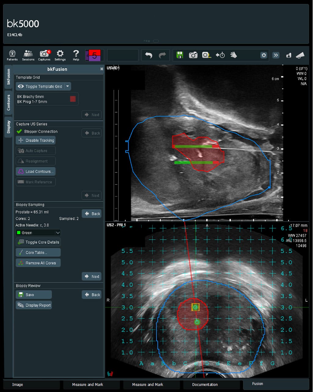
At Texas Radiotherapy, we utilize advanced MR fusion transperineal biopsy to deliver unprecedented accuracy in prostate cancer diagnosis and staging. By fusing high-resolution, pre-procedure MRI scans with real-time transrectal ultrasound imaging, this technique allows Dr. Cavey to pinpoint and biopsy suspicious lesions directly – substantially reducing the likelihood of missing aggressive, high-grade cancers or understaging tumors during active surveillance.
The MR-fusion transperineal biopsy is an improvement over the traditional transrectal ultrasound method because the MR-fusion transperineal biopsy targets specific lesions versus random sampling. Additionally, transperineal biopsies can reach parts of the prostate that are nearly impossible to reach during a transrectal biopsy. Transperineal biopsies are much “cleaner” than transrectal biopsies and have less risk of infection. Transperineal biopsies can be performed in the office with local anesthesia or in the operating room under general anesthesia. Patients have reported less discomfort with transperineal as opposed to transrectal biopsies. The MR-fusion transperineal technique is now the standard of care for prostate biopsies.
For decades, the standard transrectal ultrasound (TRUS) biopsy was the primary method used across the United States. However, traditional transrectal biopsies carry significant limitations:
Texas Radiotherapy performs MRI-TRUS fusion transperineal biopsies where MRI images are fused with the TRUS image to identify suspicious lesions for biopsy. MRI-TRUS transperineal biopsy targets specific lesions whereas TRUS uses ultrasound guidance to assist the practitioner in taking samples from multiple standard cores throughout the prostate with no specific lesion target. The MR fusion transperineal biopsy technique improves the accuracy of identifying high-grade tumors while minimizing the overdetection of low-grade tumors. Additionally, transperineal biopsies can reach parts of the prostate that are nearly impossible to reach during a transrectal biopsy.
During the transperineal biopsy, the needle passes from the skin below the scrotum into the prostate. Transperineal biopsies are much “cleaner” and are associated with a near zero percent chance of serious bacterial infection. Transperineal biopsies can be performed with sedation in an operating room or in the office with local anesthesia. In addition, patients report less discomfort with transperineal as opposed to transrectal biopsies.
The Advantages of MR fusion transperineal biopsy
By shifting to an MRI-guided transperineal approach, Texas Radiotherapy provides an advanced, safer diagnostic standard:
Traditional biopsies pass through the rectal wall, carrying a serious risk of introducing bacteria into the prostate and bloodstream. A transperineal biopsy enters through external skin, virtually eliminating the risk of sepsis while utilizing 3D MRI fusion to directly target suspicious lesions that traditional methods frequently miss.
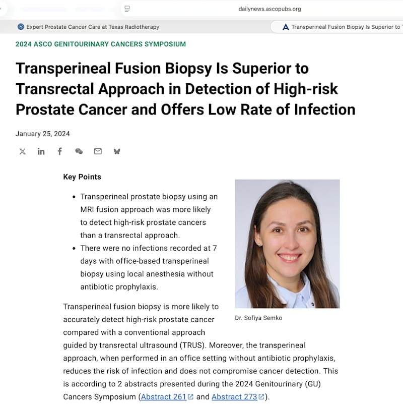
Dr. Cavey will perform these for initial confirmation, for a patient that has a PSA that continues to increase, but prior biopsies did not confirm prostate cancer, or to confirm the correct stage. MR fusion transperineal biopsy is proven to be superior to rectal biopsies.
Most patients resume normal, light daily activities within 24 to 48 hours. Because there are no punctures through the rectal lining, rectal bleeding is avoided. Minor perineal soreness or mild blood in the urine/semen can occur temporarily and typically resolves within a few days.
Depending on patient preference, anatomical factors, and clinical needs, the procedure can be performed in-office with local anesthesia or in an outpatient surgical setting under light sedation or general anesthesia.